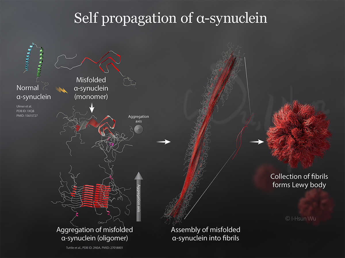
The purpose of the illustration is to teach graduate students and conference attendees the process of alpha-Synuclein self propagation on a powerpoint slide. Alpha-Synuclein, is an abundant protein whose function in the healthy brain is not well understood yet. However, widespread aggregation of the misfolded alpha-Synuclein in the form of Lewy bodies is one of neuropathological hallmarks of Parkinson’s disease. The misfolded alpha-Synuclein, rich in beta-sheet structure (shown in red) can be the precursor of aggregation. The illustration depicts steps of self propagation of alpha-Synuclein from misfolded monomers->oligomers->fibrils->Lewy bodies. Crystal structures of the normal alpha-Synuclein, monomer and oligomer of misfolded alpha-Synuclein were obtended via Protein Data Bank and then brought to Cinema 4D for finer adjustments on materials and lighting. The model of fibril and Lewy bodies were generated using Cinema 4D.
- Media: PDB, Cinema 4D, Adobe Photoshop, Illustrator CC
- Intended Audience: Graduate students, basic scientists, neurologists