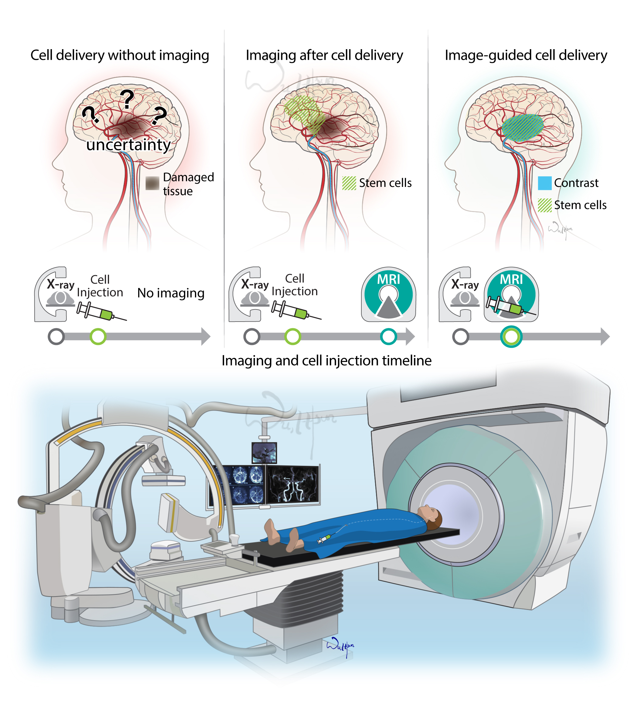
The purpose of this illustration is to promote the use of magnetic resonance imaging during stem cell infusion to target stroke damage site for optimal neuro-regeneration. Drawings on top show three scenarios of imaging usage and cell injection and their outcomes. (1) without MRI, there is uncertainty where the cell deployed and no access to tracking. (2) MRI after cell injection, the MRI readout could be a reference for the future treatment plan; however, there is no advice on targeting the injured area during cell injection. (3) MRI guided cell injection, the cell deployment is matched with the targeted area due to the real-time MR imaging visualization. The illustration at the bottom shows the setting of the MRI system (on the right) and the X-ray system (on the left) in an angiography suite. This illustration is one of a series of illustrations in the paper “Intra-Arterial Delivery of Cell Therapies for Stroke,” published in the May 2018 issue of Stroke
- Media: Adobe Illustrator CC
- Intended Audience: Radiologists, neurologists